Tumor Imaging
CPN’s high brightness, photostability, and nontoxicity are promising for live cell/tissue imaging (Angew. Chem., 2007). We are interested in developing CP-based biomaterials for tumor specific imaging and therapeutic delivery by functionalizing CPs with targeting ligands and biopolymers. Collaborating with Prof. Peter So at MIT, we demonstrated that CPNs are extremely bright and stable two-photon (2P) materials exhibiting nontoxicity.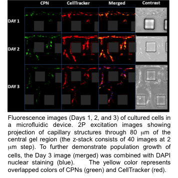
Using a tissue culture model, we imaged endothelial cell growth up to three days without observing any toxic effects (Adv. Mater., 2009). By complexing CPNs with cancer cell specific hyaluronic acid (HA), we demonstrated cancer cell-specific labeling with low binding to normal cells (Macromolecules, 2013). We are synthesizing biodegradable CPN/HA hybrid materials for in vivo tumor labeling and drug/gene delivery. We are investigating how the aggregation structure influences 2P cross-section and tumor specific imaging using temporal focusing 2P excitation with Prof. So at MIT. The CPNs’ in vivo properties are under testing in a mouse model system. NIH funding was received to develop 2P-imaging probes and to test target cancer cell specificity in vitro. We are collecting in vivo pharmacokinetic and toxicology data of CPNs and CPN/HA in collaboration with prof. McGoron in the Biomedical Engineering Department at FIU.